


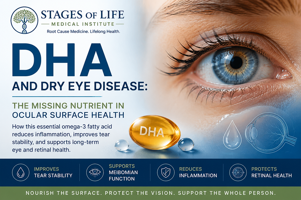
DHA and Dry Eye Disease: The Missing Nutrient in Ocular Surface Health

Quick Look
Dry eye disease affects millions of adults, yet treatment often focuses only on symptom relief. Increasing evidence suggests that nutritional deficiencies — particularly low intake of the omega-3 fatty acid docosahexaenoic acid (DHA) — may contribute significantly to chronic ocular inflammation, unstable tear production, and progressive surface irritation. Supporting ocular tissues nutritionally may represent an overlooked therapeutic opportunity.
Dry Eye Disease Is Not Simply a Lubrication Problem
Patients frequently describe symptoms of:
Burning
Eye fatigue
Intermittent blurred vision
Foreign body sensation
Excess tearing
Light sensitivity
Difficulty wearing contact lenses
Conventional treatment usually involves artificial tears or prescription anti-inflammatory drops. Unfortunately, these approaches often address symptoms while overlooking a deeper issue.
Dry eye disease increasingly appears to represent a disorder of chronic ocular surface inflammation, tear film instability, oxidative stress, and dysfunction of the meibomian glands.¹²
For many patients, nutrition may be part of the solution.
Why DHA Matters
Docosahexaenoic Acid (DHA) is an essential omega-3 fatty acid highly concentrated within the eye.
DHA is found primarily in:
Retinal photoreceptors
Corneal epithelial cell membranes
Neural tissues
Meibomian gland lipid secretions
Nearly 50% of retinal membrane phospholipids contain DHA, making it one of the most concentrated fatty acids in ocular tissue.³
This is not accidental. Vision depends upon membrane flexibility, proper cell signaling, and control of inflammation.
Understanding the Tear Film
Healthy vision depends on a highly organized tear film consisting of three separate layers.
Lipid Layer
Produced by the meibomian glands; reduces evaporation.
Aqueous Layer
Produced by lacrimal glands; provides hydration.
Mucin Layer
Allows tears to adhere uniformly across the corneal surface.
Failure of any layer destabilizes the entire system.
This produces increased evaporation, irritation, inflammation, and progressive epithelial injury.

Four Mechanisms Through Which DHA Supports Dry Eye Improvement
1. DHA Reduces Inflammation
DHA serves as precursor for a family of powerful anti-inflammatory mediators known as:
Specialized Pro-Resolving Mediators. These compounds actively terminate inflammation.
They reduce:
TNF-alpha
IL-1 beta
Matrix metalloproteinase activity
Corneal inflammatory injury⁴
Unlike conventional medications, these molecules help resolve inflammatory signaling naturally.
2. DHA Supports Meibomian Gland Function
Meibomian Gland Dysfunction remains the most common cause of chronic dry eye.
The lipid secretions from these glands require healthy fatty acid composition.
Reduced DHA availability may impair lipid quality and accelerate tear evaporation.⁵
3. DHA Stabilizes Cellular Membranes
Healthy ocular tissues require membrane fluidity.
DHA helps maintain structural integrity within:
Corneal epithelial cells
Conjunctival tissues
Lacrimal gland membranes
Neural signaling pathways within retinal tissues
Poor membrane fluidity predisposes tissues to oxidative injury.
4. DHA Protects the Retina
Beyond dry eye itself, DHA protects deeper retinal tissues against oxidative stress and may reduce progression of age-related degeneration.⁶
Thus, DHA simultaneously supports:
Ocular comfort
Surface healing
Retinal longevity

Clinical Pearl
One of the overlooked associations in chronic dry eye disease is the presence of broader systemic inflammation.
Patients frequently demonstrate concurrent:
Insulin resistance
Hyperglycemia
Autoimmune activation
Hormonal imbalance
Poor omega-3 intake
Diets dominated by processed seed oils
The eyes may simply be the first organ signaling inflammatory dysfunction occurring elsewhere.
Dry eye disease deserves a broader metabolic evaluation.
Who Should Consider DHA Support?
Higher-risk groups include:
Adults over age 50
Post-menopausal women
Patients with autoimmune disease
Heavy computer users
Contact lens wearers
Patients with chronic inflammatory disorders
Individuals with Sjögren Syndrome
Individuals consuming low marine-fat diets
Dietary Sources of DHA
Highest natural sources include:
Salmon, Sardines, Mackerel, Anchovies
Plant-derived omega-3 fats convert poorly to DHA.
Many patients may require direct supplementation.

While dietary intake from cold-water fish remains ideal, many individuals do not consume sufficient amounts of omega-3 fatty acids to achieve therapeutic benefit. For patients with chronic dry eye symptoms, most clinical studies evaluating ocular surface improvement have used combined omega-3 supplementation in the range of 1,000–3,000 mg daily (Total of all Omega 3's), with evidence suggesting that formulas providing at least 500–1,000 mg of DHA. per day.

Docosahexaenoic Acid (DHA) may offer particular benefit for tear film stability, meibomian gland support, and reduction of ocular inflammation. Higher doses may be appropriate in selected individuals, especially when systemic inflammation, autoimmune disease, or poor dietary intake are present. As with any supplement, purity, oxidation stability, and third-party quality testing remain critically important when selecting an omega-3 formulation.⁷⁸
Related Topics You May Find Helpful
Dry eye disease rarely exists in isolation. Ocular inflammation, membrane instability, oxidative stress, and poor nutritional intake often reflect broader systemic health concerns. Understanding these related conditions can provide important insight into the underlying causes of chronic symptoms.
If this topic interests you, you may also benefit from reading:
1. Pre-Diabetes: Is It a Real Thing?
Elevated blood sugar and insulin resistance contribute directly to systemic inflammation, vascular dysfunction, and accelerated tissue damage throughout the body — including the delicate microvascular structures of the eye. Blog to be Published Soon!
Bottom Line
Dry eye disease should no longer be viewed simply as inadequate lubrication.
It increasingly appears to represent a disorder involving:
Chronic inflammation
Tear film instability
Meibomian gland dysfunction
Oxidative stress
Nutritional insufficiency
DHA is one of the most structurally important fats in the human eye.
Artificial tears provide temporary comfort. Correcting nutritional deficiency may provide lasting improvement.
Become a Patient
At Stages of Life Medical Institute, we focus on identifying root causes of chronic disease using advanced diagnostics, nutritional science, hormone evaluation, and physician-guided integrative medicine.
Persistent symptoms often deserve deeper answers.
At Stages of Life Medical Institute, we believe symptoms rarely occur in isolation. The body operates as an integrated system — and identifying root causes often reveals connections conventional medicine overlooks.
References
1. Miljanović B, Trivedi KA, Dana MR, et al. Relation between dietary n-3 and n-6 fatty acids and clinically diagnosed dry eye syndrome in women. American Journal of Clinical Nutrition. 2005;82(4):887-893.PubMed:PubMed Reference 1
2. Craig JP, Nichols KK, Akpek EK, et al. TFOS DEWS II Definition and Classification Report. Ocular Surface. 2017;15(3):276-283.PubMed:PubMed Reference 2
3. Bazan NG. Cell survival matters: docosahexaenoic acid signaling, neuroprotection and photoreceptors. Trends in Neurosciences. 2006;29(5):263-271.PubMed:PubMed Reference 3
4. Rashid S, Jin Y, Ecoiffier T, et al. Topical omega-3 fatty acids and corneal inflammatory response in dry eye models. Investigative Ophthalmology & Visual Science. 2008;49(2):652-661.PubMed:PubMed Reference 4
5. Calder PC. Omega-3 fatty acids and inflammatory processes: from molecules to man. Nutrients. 2017;9(9):E1016.PubMed:PubMed Reference 5
6. Eom Y, Lee JS, Kang SY, et al. Omega-3 fatty acid supplementation and meibomian gland dysfunction. Cornea. 2016.PubMed:PubMed Reference 6
7. Bhargava R, Kumar P, Arora Y. Oral omega-3 fatty acid treatment in dry eye syndrome. Cornea. 2013;32(10):1297-1304.PubMed:PubMed Reference 7
8. Downie LE, Keller PR. Systematic review of omega-3 interventions for dry eye disease. Ocular Surface. 2015.PubMed:PubMed Reference 8
9. Nichols KK, Foulks GN, Bron AJ, et al. TFOS DEWS II Pathophysiology Report. Ocular Surface. 2017.PubMed:PubMed Reference 9
10. Clayton JA. Dry eye disease pathophysiology and modern treatment strategies. JAMA. 2018.
The medical references cited in this article are provided for educational purposes only and are intended to support general scientific discussion. They are not a substitute for individualized medical advice, diagnosis, or treatment. Clinical decisions should always be made in consultation with a qualified healthcare professional who can account for a patient’s unique medical history, medications, and circumstances.


1917 Boothe Circle, Suite 171
Longwood, Florida 32750
Tel: 407-679-3337
Fax: 407-678-7246



.webp)